Medical Chest Bucky Stand: Definition, Structural Analysis & Clinical Applications
Author:
Technical Overview: Chest Bucky Stand (Upright Radiographic Stand)
The chest bucky stand—formally known as an upright radiographic stand or wall bucky stand—is a core auxiliary device in medical X-ray imaging systems . It is designed to support patients in standing or sitting positions for radiographic examinations of the chest, skull, abdomen, pelvis, and other anatomical regions .

1. Medical Definition & Core Functions
A chest bucky stand is a vertically adjustable imaging accessory that pairs with various X-ray generators. Its primary functions include:
- Patient Stabilization: The upright column structure supports the patient, ensuring body stability during exposure to minimize motion blur.
- Scatter Radiation Filtration: Integrated high-density grids (grid density ≥ 40 lines/cm) effectively absorb scattered radiation, enhancing image contrast .
- Multi-Region Adaptability: Supports imaging from the skull (orbital-meatal line level) to the abdomen (seated AP view), with a vertical travel range of 1000–1830mm .
Modern chest stands universally employ grid-equipped configurations, with grid ratios typically 10:1 or higher for high-kV chest radiography .

2. Structural Composition & Technical Parameters
Structural Breakdown:
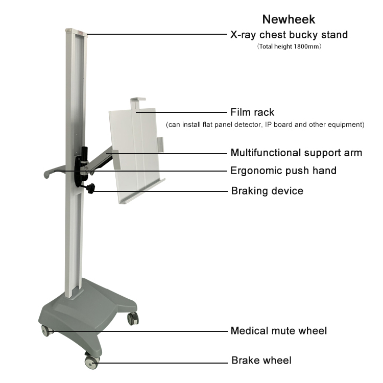
- Column System:
- Constructed from high-strength aluminum alloy. Some models feature floor-mounting fixtures or mobile casters (for mobile versions, caster material and locking stability are critical).
- Wall-mounted stands require wall load-bearing capacity ≥ 150kg, with installation height ≥ 500mm from the floor.
- Carriage Mechanism:
- Integrated with auto-locking functionality, anti-collision sensors, and emergency stop buttons.
- Vertical travel deviation ≤ ±5mm; operating force ≤ 50N.
- Supports both manual and motorized dual-mode control.
- Cassette Assembly:
- Houses a removable grid (grid ratio 8:1 to 12:1), adaptable to different kV conditions .
- Compatible with detector sizes from 5″×7″ up to 17″×17″, supporting DR flat panel detectors, CR IP plates, and traditional film cassettes .
Key Technical Parameters:
| Parameter | Specification |
|---|
| Vertical Travel | ≥ 1500mm (detector center to floor) |
| Grid Focal Distance | 180cm (matched to tube distance) |
| Grid Ratio | 10:1 or 12:1 (for chest dedicated stands) |
| Electrical Safety | Ground resistance ≤ 0.1Ω; insulation resistance ≥ 20MΩ |
| Radiation Protection | Built-in lead curtain (lead equivalent ≥ 0.5mmPb) |
3. Clinical Application Scenarios
Chest Imaging:
- Stabilizes standing patients for PA and lateral thoracic views, reducing blur caused by respiratory motion.
- Paired with Automatic Exposure Control (AEC), ionization chamber response time ≤ 100ms.
Specialized Region Imaging:
- Skull radiography: Carriage adjusts to orbital-meatal line level; paired with cephalometric fixators for temporal bone/sinus examinations.
- Abdominal imaging: Accommodates seated patients; cassette height is adjusted via the lift mechanism.
Emergency & Mobile Scenarios:
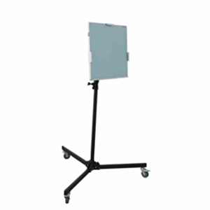
- Mobile chest stands equipped with casters and locking devices are suitable for ICU or bedside radiography, though load capacity is typically ≤ 150kg.
4. Classification & Selection Criteria
By Mobility:
- Fixed Upright Chest Stand: Permanent installation, load capacity ≥ 200kg. Ideal for high-throughput radiology departments.
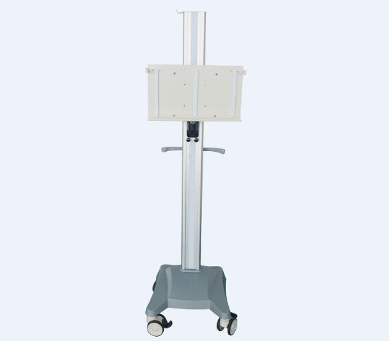
- Mobile Chest Stand: Equipped with nylon + metal bearing casters. Full-load locking stability must be validated during testing.
By Detector Type:
- CR Cassette Type: Compatible with traditional cassettes and IP plates.
- DR Flat Panel Type: Supports wireless flat panel detector direct charging (contact impedance ≤ 0.5Ω).
Brand & Compliance:
- Prioritize manufacturers certified under ISO 13485. Warranty periods should cover critical components such as motors and grids.
💡 Key Procurement Insights for International Markets:
Based on industry standards and clinical requirements, here are the critical decision factors for overseas buyers:
- Grid Configuration is Paramount: For chest-dedicated stands, specify a 10:1 or 12:1 grid ratio with 180cm focal distance . This is the international benchmark for optimal scatter rejection in chest radiography.
- Detector Compatibility: Ensure the cassette assembly accommodates 17″×17″ wireless DR detectors—the current global standard for digital radiography .
- Travel Range Verification: A minimum vertical travel of 1100mm is the industry baseline , but premium models extend to 1500mm+ for full-body upright imaging.
- AEC Integration: For departments upgrading to modern DR systems, confirm the stand supports 3-chamber AEC integration for consistent dose optimization .
Summary
The chest bucky stand is far more than a simple support frame—it is a precision optical-mechanical assembly that directly impacts diagnostic image quality. When selecting a unit, prioritize grid ratio (10:1+), vertical travel range (≥1100mm), detector compatibility (up to 17″×17″ DR panels), and manufacturer certifications (ISO 13485). Whether for a fixed installation in a high-volume radiology suite or a mobile unit for ICU bedside exams, the right chest stand ensures both patient safety and superior imaging outcomes.
FAQ:
Q: What is a chest bucky stand used for?
A chest bucky stand holds the cassette or digital detector upright during chest radiography. The patient stands in front of it, and the bucky grid removes scattered radiation so the lungs and ribs appear sharp on the image.
Q: What is the difference between a chest bucky and a wall bucky?
The terms are often used interchangeably. A chest bucky emphasizes the application (chest radiography), while a wall bucky emphasizes the mounting method. Most chest bucky stands sold today are wall-mounted vertical stands.
Q: Which chest bucky stand is best for a small clinic?
For small clinics, a wall-mounted chest bucky stand with a 10:1 grid and 14″ × 17″ film box is the most practical choice — it saves floor space and covers all routine chest examinations.
