Vertical Stitching Radiography Stand for Full-Spine and Long-Bone Imaging: Technical Parameters, Core Designs, and Clinical Applications
When conducting long-length radiography such as full-spine scoliosis screening and weight-bearing full-leg imaging, radiology departments often face three major pain points: bulky equipment footprints, image misalignment at the stitching seams, and time-consuming, labor-intensive positioning for radiographers.
This Vertical Stitching Radiography Stand is engineered to solve these exact clinical challenges. With its rigid mechanical structure and smooth sliding rail system, it seamlessly integrates with standard DR systems and stitching software. It delivers high-precision, distortion-free image stitching even in compact imaging rooms.
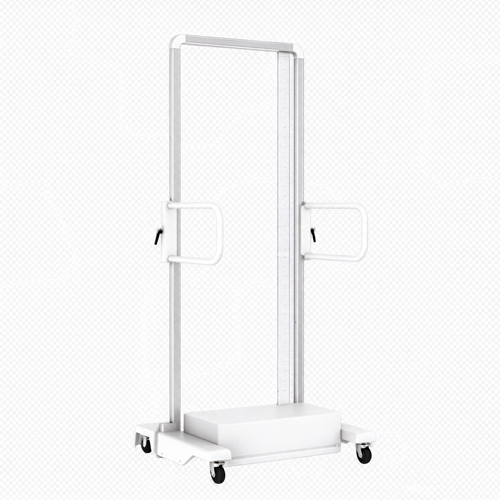
Below is a detailed breakdown of its core parameters, structural features, and clinical applications:
I. Core Specifications (First Priority for Equipment Selection)
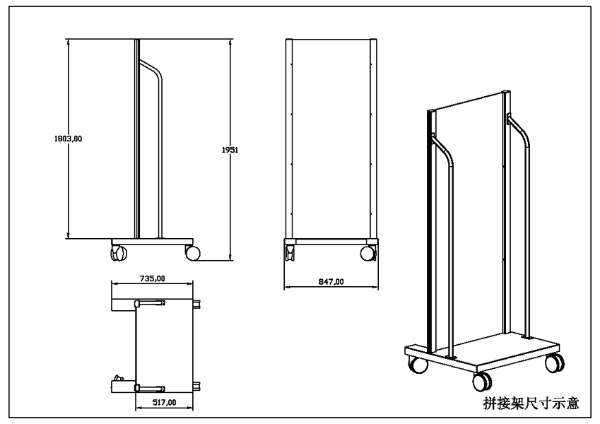
Based on the engineering drawings, the stand features ergonomic dimensions that perfectly balance patient accommodation and compact room layout:
- Total Height: 1985 mm (Accommodates full-spine and full-lower-limb scans for the vast majority of adults)
- Total Width: 967 mm (Provides stability without excessive space consumption)
- Max Depth: 725 mm (Including the step and base extension)
- Rail/Main Body Depth: 517 mm (Low-profile design, suitable for various compact DR rooms)
- Structure Material: High-strength metal frame (ensures no deformation under long-term load)
- Installation: Floor-standing/wall-mounted, compatible with ceiling-suspended and floor-mounted X-ray tube systems
II. Core Features and Structural Advantages
- High-Rigidity Load-Bearing Frame to Eliminate Micro-Vibrations
- The integrated reinforced column structure provides exceptional stability when patients hold onto it. During multiple consecutive exposures (the stitching process), it effectively eliminates image gaps and artifacts caused by frame deformation or subtle shaking, ensuring sub-millimeter stitching accuracy.
- Low-Resistance Sliding Rail for Effortless Operation
- Equipped with a precision sliding mechanism, the detector tray moves up and down smoothly without jamming. Combined with a user-friendly locking handle, radiographers can complete positioning quickly and accurately, significantly reducing daily workload and physical exertion.
- Compact Base Design to Maximize Room Space
- The overall depth is controlled within 725 mm, with the rail section at only 517 mm. The retracted base design (as shown in the drawings) ensures it does not interfere with standard chest or table radiography when not in use for long-length exams.
- Wide Anti-Slip Step and Multi-Position Adaptability
- Features a stable standing base where patients can plant their feet naturally to ensure a true weight-bearing state during full-leg radiography. Handrails on both sides of the column help scoliosis or elderly patients maintain a stable posture, reducing motion artifacts.
III. Clinical Application Scenarios
This stand is not just a physical support but an “accuracy benchmark” for complex long-field imaging. It is primarily used in the following clinical fields:
- Orthopedics / Spine Surgery:
- Full-Spine AP/Lateral Stitching: For precise Cobb angle measurement in scoliosis, and pre-/post-operative evaluation of kyphosis and lordosis.
- Full-Leg Stitching: For assessing varus/valgus knee deformities and leg-length discrepancy (LLD), providing accurate mechanical axis data for joint replacement and osteotomy surgeries.
- Sports Medicine and Rehabilitation:
- Weight-bearing/non-weight-bearing comparative radiography of the full spine and pelvis to evaluate postural changes before and after rehabilitation.
- Occupational Health (e.g., Pneumoconiosis):
- Large-field spine examinations required for specific physical checkups.
IV. Seamless Integration with Existing DR Systems
- Precise Synchronization: Adopts a universal mechanical interface, perfectly matching the automatic tube-to-detector tracking features of mainstream DR systems.
- Plug-and-Play: Works with the department’s existing DR workstation and auto-stitching algorithms, eliminating tedious manual alignment and rapidly outputting distortion-free, high-resolution long-field X-ray images.
Conclusion:
With 1985 mm of vertical coverage, a high-rigidity anti-vibration frame, and a rail depth of just 517 mm, this vertical stitching radiography stand achieves the perfect balance between “space efficiency” and “diagnostic accuracy.” It is an ideal hardware foundation for radiology departments looking to upgrade their long-bone and full-spine imaging capabilities.
Why Buy Direct from Newheek?
ISO 13485Certified Quality Management System
CE CertifiedCompliant products for global markets
Factory-Direct16-year manufacturer, no middleman
Global ShippingExperienced export & delivery support
