X-Ray Bucky Stand: The Unsung Hero of Chest Radiology & Imaging Precision
When you look at a chest X-ray, your eyes naturally gravitate toward the lung markings or the silhouette of the heart. You likely never consider that the primary “unsung hero” behind that crystal-clear image is a simple, stationary structure standing quietly in the corner of the radiology room: the X-ray Bucky Stand.
In the shadow of high-tech giants like CT and MRI scanners, the vertical Bucky stand can seem overly “basic.” However, from a radiologist’s perspective, the quality and calibration of this device directly dictate whether a routine chest X-ray becomes a reliable diagnostic tool or a missed opportunity.
1. More Than Just a “Stand”: The Precision of the Bucky System
To the untrained eye, it’s a metal frame. To a technician, it is a masterclass in stability and positioning. A high-quality Bucky stand incorporates several critical engineering features:
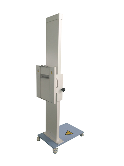
- Vibration Control: Stability is the lifeblood of imaging. The base must be heavy and the column rigid. Even a microscopic tremor during the fraction of a second when the X-rays are emitted can cause motion blur, much like a shaky hand ruins a photograph. In radiology, a “re-take” means unnecessary radiation exposure for the patient and reduced workflow efficiency.
- The “Invisible Ruler”: The distance and alignment between the Bucky tray (which houses the digital detector) and the X-ray tube are strictly standardized. This geometric relationship ensures minimal image distortion, making measurements of heart size or lesion locations accurate and trustworthy.
- Ergonomics and Human-Centric Design: Modern stands are fine-tuned for fluid movement and rotation. Technicians adjust these stands hundreds of times a day; a clunky design increases the risk of repetitive strain injuries. Furthermore, wide adjustable ranges allow for more compassionate care when imaging patients in wheelchairs or those with limited mobility.
2. From Iron Frames to Smart Nodes: The Evolution of the Bucky
The history of the Bucky stand is a microcosm of medical imaging progress.

- The Mechanical Era (1.0): Pure metal structures designed for one task: holding heavy intensifying screens and film cassettes. They were reliable but cumbersome.
- The Digital Radiography (DR) Leap (2.0): This was the most significant shift. The film tray was replaced by a digital flat-panel detector. The stand became a critical link in a digital chain, protecting equipment worth hundreds of thousands of dollars while facilitating high-speed data transfer to diagnostic workstations.
- The Intelligence Frontier (3.0): Latest-generation stands now integrate sensors and motorized drives for:
- Auto-Tracking: As the X-ray tube moves, the Bucky stand automatically aligns itself, eliminating manual adjustments.
- One-Click Positioning: Input the patient’s height, and the stand glides to the preset elevation.
- Anti-Collision Systems: Sensors detect proximity to the patient, preventing accidents and enhancing safety.
3. Why It Remains Irreplaceable
Despite the rise of 3D imaging, the standing Posteroanterior (PA) chest X-ray remains the gold standard for clinical screening and initial diagnosis.

- Supreme Efficiency: Exposure takes milliseconds. The entire process, from positioning to the final image, takes just minutes—unmatched in emergency or mass-screening scenarios.
- Cost-Effectiveness: The cost is significantly lower than a CT scan, yet it provides a wealth of information regarding pneumonia, tuberculosis, fractures, and cardiac enlargement.
- Lower Radiation Dose: The effective dose is much lower than cross-sectional imaging, making it the preferred choice for regular health monitoring and follow-ups.
4. Radiology Trivia: Behind the Bucky
Why do we usually stand for a chest X-ray?
Standing allows the diaphragm to drop, letting the lungs expand fully to show the maximum pulmonary field. It also allows gravity to reveal air-fluid levels, which is crucial for identifying conditions like pneumothorax or pleural effusion.
What is the “wall” behind the Bucky stand?
It isn’t a standard wall. It typically contains lead shielding or lead-lined materials designed to absorb residual X-rays that pass through the patient and the stand, protecting surrounding staff and equipment from scatter radiation.
Final Thoughts
In the narrative of medical progress, we often celebrate the complexity of AI and the precision of robotic surgery. Yet, we must not overlook the “anchors” of modern healthcare. The X-ray Bucky stand is a silent guardian—it doesn’t speak, but it carries the weight of life-saving data every single day. The next time you see one in a radiology department, you might view this quiet sentinel with a bit more respect.